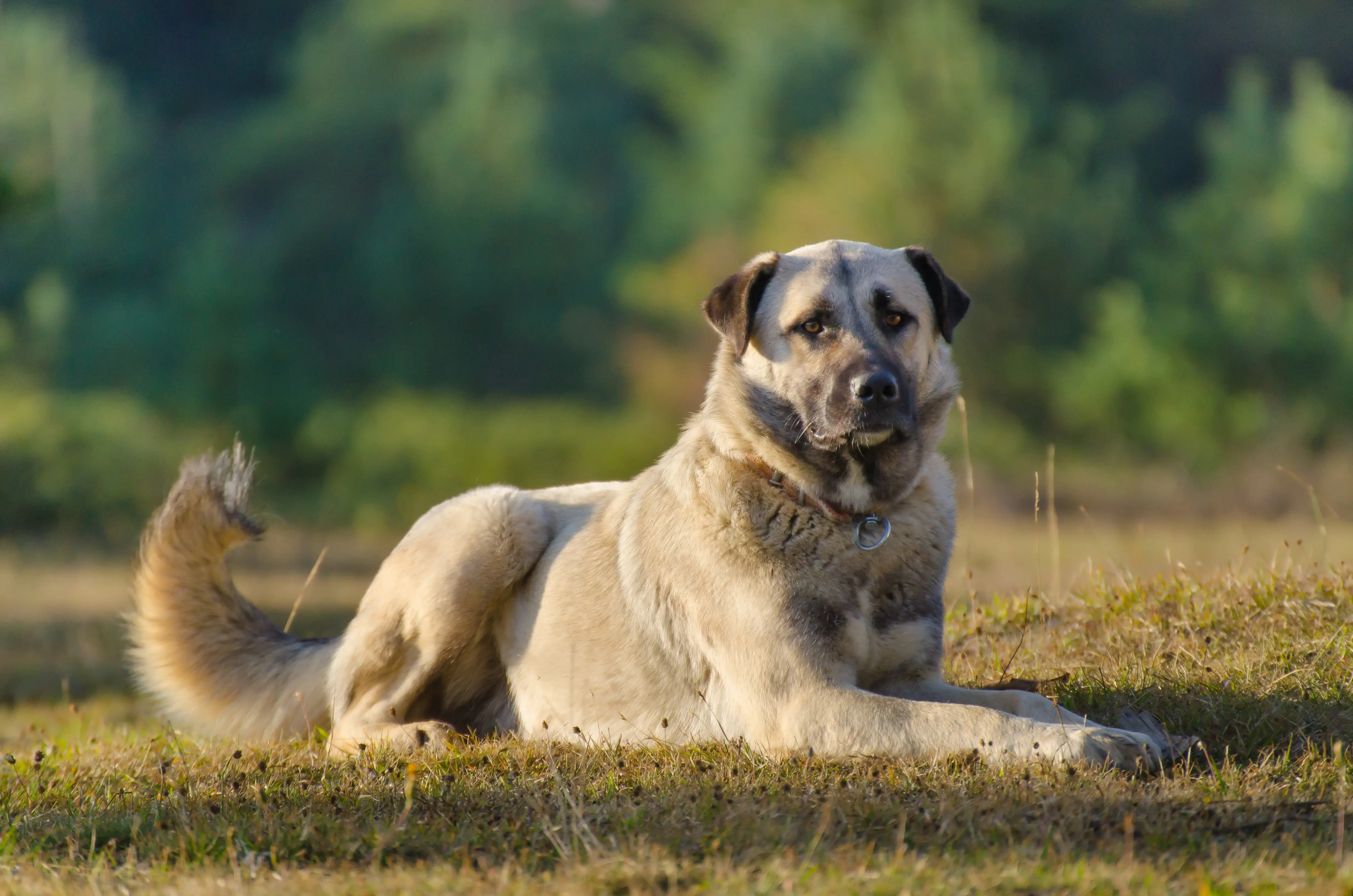
The Use of Infrared Thermography to Detect the Stages of Estrus Cycle and Ovulation Time in Anatolian Shepherd Dogs
Published by
Digatherm
on
Abstract: The aim of the study is to evaluate the effectiveness of thermographic monitoring, using the temperature changes of perianal and perivulvar areas for the determination of estrus in Anatolian Shepherd bitches. Fifteen bitches were used in the study. Blood and vaginal smear samples were collected and thermographic monitoring of perianal and perivulvar areas were carried out starting from proestrus to early diestrus.
Also, external signs of estrus were investigated. Smear samples were evaluated by light microscopy after Diff-Quik staining method and superficial and keratinized superficial cells were determined as percentage (S + KS%). Progesterone and luteinizing hormone measurements were done by radioimmunoassay. The difference in temperature between perianal and perivulvar areas was evaluated through thermographic images by FLIR ResearchIR Software.
Results: According to the results obtained from the study, differences between progesterone and S + KS% were statistically significant (P < 0.05). Although temperature showed increase and decrease with progesterone and S + KS%, the differences were not important statistically (P > 0.05). Serum luteinizing hormone levels did not sign any difference (P > 0.05).
Conclusions: As a result, thermographic monitoring alone is not enough for estrus detection in Anatolian Shepherd bitches. However, it can be used to assist the actual estrus detection technique in terms of providing some foreknowledge by evaluating the differences in temperature.
Reference: Kemal Tuna Olğaç, Ergun Akçay, Beste Çil , Burak Mehmet Uçar, Ali Daşkın (2017) J Anim Sci Technol
| Interested in learning more about thermal imaging? Request a demonstration with Digatherm and discover how veterinary thermography can help you find problem areas faster and easily monitor treatment progress. |