Abstract: Infrared thermography is a non-invasive and non-ionizing imaging technique for recording body surface temperature. Considerable research efforts have been made to ensure that thermography in human medicine is performed to consistent standards so that measurements are traceable, reliable and valid, but interest in understanding sources of measurement uncertainty has developed less rapidly in veterinary thermography.
The objective of our study was to understand the influence of variability in thermal imager performance, and the contribution to measurement variance of operator-dependent factors, in a practical equine thermography setting. The study employed five different models of thermal imager, which were all quality assured against a blackbody source prior to equine measurements.
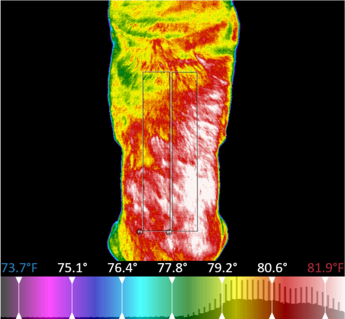
Three standard thermographic views were then obtained from nine clinically healthy horses using all five thermal imagers, with each imager capturing all views twice. On every thermographic image, regions of interest (ROIs) were then determined by two analyzers. The thermograms from each imager were assessed for their clinical utility. Agreement between analyzers of the same images, and between pairs of repeated images for the same analyzer, was assessed by Bland Altman plots.
The contribution to the total variance in temperature measurements from analyzers and from camera-dependent factors was evaluated by ANOVA gauge R&R (reliability and reproducibility) analysis. Two of the thermal imagers produced readings during quality assurance which were outside of their stated specifications for accuracy. There were notable differences in the ability of the imagers to produce clinically useful thermograms, and it was difficult to compare the thermograms from different manufacturers due to the varied color palettes employed.
Assessing agreement between analyzers, the bias was −0.05 °C and limits of agreement were 0.11 °C (upper) and −0.21 °C (lower). Agreement between repeated images was less good than between analyzers: the bias was 0.17 °C and limits of agreement were 1.55 °C (upper) and −1.21 °C (lower) for the first analyzer. Across all horses, the contribution by the analyzers to variance in temperature measurement at the ROIs from the lateral whole-body view was 9.8 } 4.2% (Mean } SD), for the lateral and medial aspects of the limbs was 26.5 } 18.2%, and for the dorsal aspect of the limbs was 11.2 } 9.2%. Across all ROIs and horses, the mean contribution to variance in the measurements from camera factors was 82.7 } 15.3%. The work shows the importance of specifying an appropriate thermal imager for equine studies, and ensuring there is a programme of quality assurance in place for all devices.
Reference: Kevin Howell, Krzysztof Dudek, Maria Soroko (2020). Infrared Physics and Technology 110 103447
|
Interested in learning more about thermal imaging? Request a demonstration with Digatherm and discover how veterinary thermography can help you find problem areas faster and easily monitor treatment progress.
|