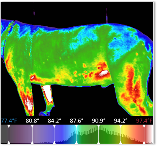
Abstract: This paper work reviews the main clinical and imaging diagnostic methods of cranial cruciate ligament rupture in dogs and shows the results of using Flir E4 thermal imaging camera and Flir tools 2017 software analysis.
Compared to the achievements of a correct cranial cruciate ligament ruptures diagnosis in dogs of 75-85% reported by Infemuso et al, 2010 using Med 2000 IRIS type thermographic camera, Meditherm Inc with intranet service for image interpretation and analysis with Automated Computer Vision software and Image processing-Algorithm Test and Analysis Tool (CVIP-ATAT) data obtained by us with Flir E-40 thermal imaging camera and Flir tools-2017 analysis software did not allow the recognition of one pattern for differentiation between dogs with healthy cranial cruciate ligament (CCL) and with cranial cruciate ligament rupture (CCLR) as well as with knee osteoarthritis (OA). Thermal imaging camera and tested software allows differentiation of dogs without knee pathology by those with injured knees, without revealing significant differences among different pathological entities (CCLR versus OA).
Reference: Igna, C., Mavromatis, S., Bumb, D., Sicoe, B., Zaha, C. & Schuszler, L. (2017). Thermal Imaging of the Dogs with Cranial Cruciate Ligament Ruptures. VOL. L.
|
Interested in learning more about thermal imaging? Request a demonstration with Digatherm and discover how veterinary thermography can help you find problem areas faster and easily monitor treatment progress.
|