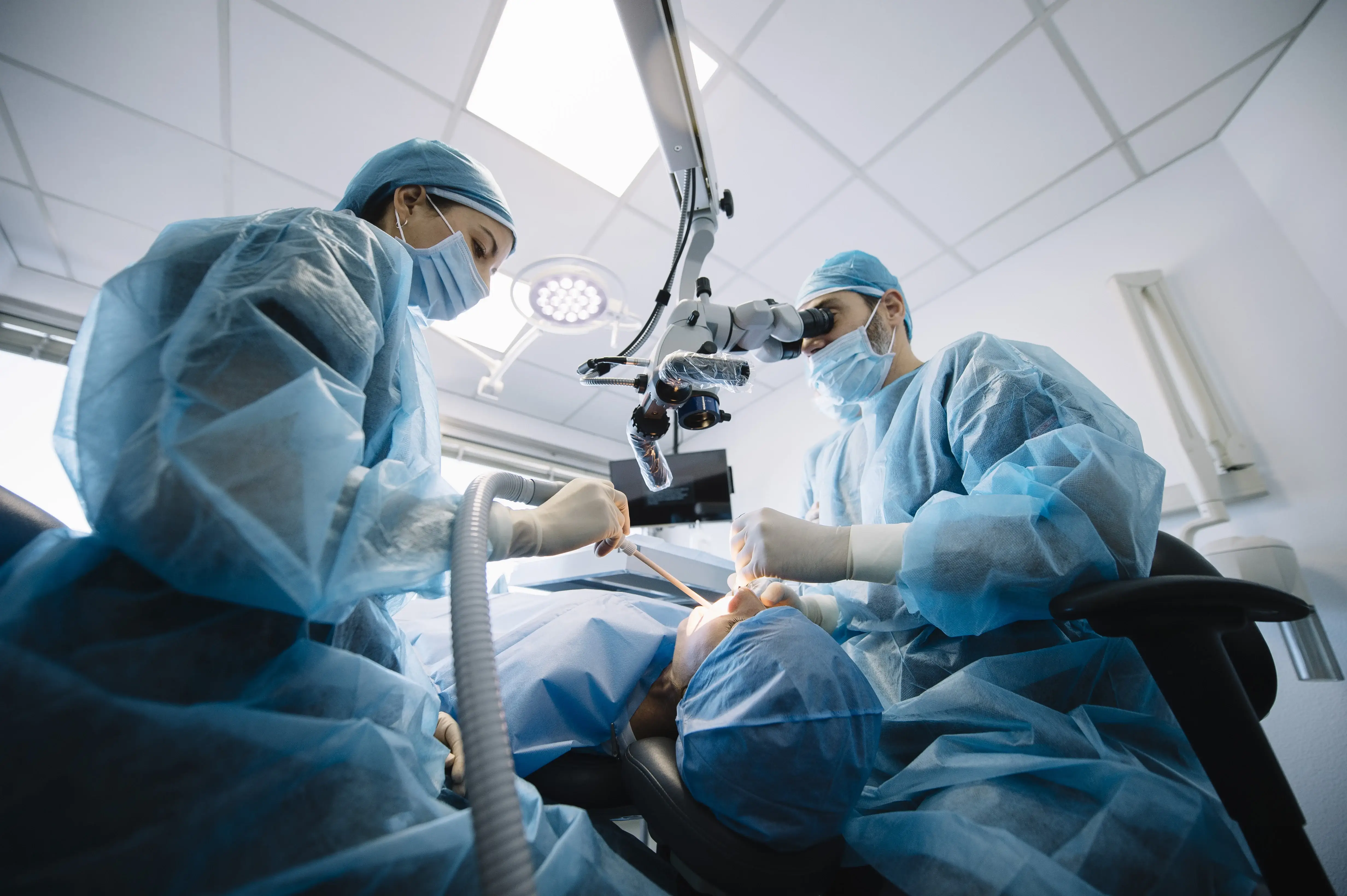
Medical Infrared Thermography in Peri-Operative Management of Peripheral Ameloblastoma
Abstract: Peripheral ameloblastoma (PA) is a rare benign peripheral odontogenic tumor arising in the gingiva and in the overlying mucosa of tooth-bearing areas of the jaws. Recent data suggestthat the recurrence rate is directly related to inadequate surgical excision This case of a 71-year-old man reports a poorly delineated mass effecting the gum of the left mandibular caninepremolars area histologically corresponded to PA. In complement to clinical visual examination of such a poorly delineated, non-exophytic and non-dyschromic inflammatory lesion, medical infrared thermography (MIT) - a non-invasive, non-ionizing and real-time imaging technique - was used to optimize the soft tissue margins, and a marginal bone resection was performed. MIT has also been found to be a useful tool in monitoring the absence of diseased tissue crossing the excisional margins at the end of the operation to minimize the risk of recurrence. After two years of follow-up, no local recurrence was found.
Reference: Maxime Delarue, Stéphane Derruau , Paul Troyon , Fabien Bogard , Guillaume Polidori, Cédric Mauprivez (2021) Photodiagnosis Photodyn Ther Jun;34:102167
| Interested in learning more about thermal imaging? Request a demonstration with Digatherm and discover how veterinary thermography can help you find problem areas faster and easily monitor treatment progress. |

