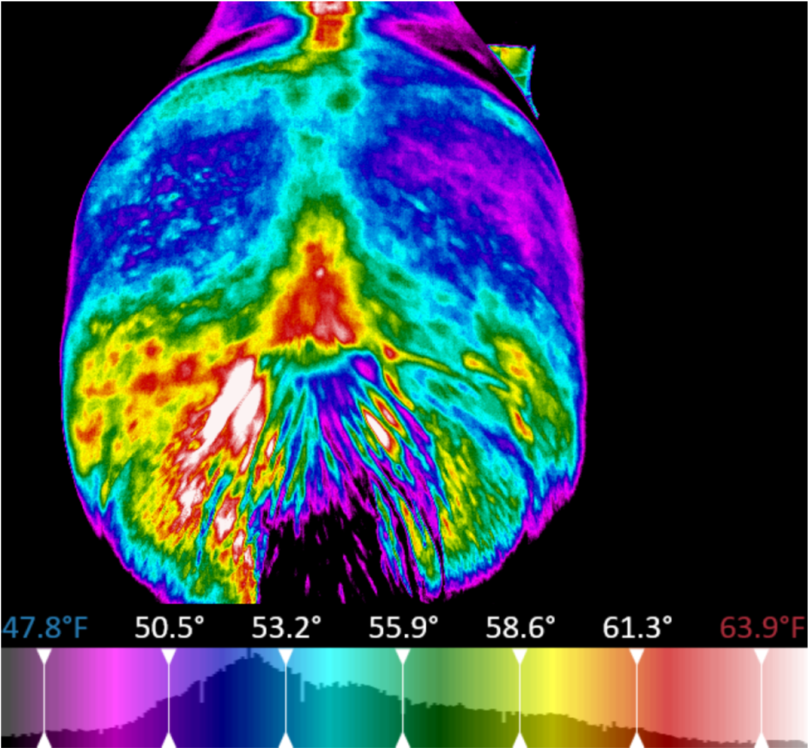
Thermography as an Aid to the Clinical Lameness Evaluation
Abstract: Thermography has been shown to be a practical aid in the clinical evaluation of lameness. This modality specifically increases the accuracy of diagnosis. Thermography represents skin temperature, usually pictorially.
The techniques involve contacting and noncontacting modalities. Noncontacting thermography, which detects infrared radiation, is the most accurate. In order to be accurate, thermography must be performed in a temperature controlled, draft-free area. The area should be protected from sunlight to avoid erroneous heating of the skin, and the hair length should be uniform.
Thermography detects heat before it is perceptible during routine physical examination; therefore, it is useful for early detection of laminitis, stress fractures, and tendinitis. It offers a noninvasive means of evaluating the blood supply to an injured part and offers one of the only reliable means to evaluate blood flow to the foot of horses with navicular syndrome.
Thermography also is useful for the early identification of stress injuries to the contralateral limb of convalescing orthopedic patients. Thermography is an excellent adjunct to clinical and radiographic examination. It is complementary to other imaging techniques such as ultrasonography and scintigraphy.
Reference: T A Turner. (1991) Vet Clin North Am Equine Pract Aug;7(2):311-38
|
Interested in learning more about thermal imaging? Request a demonstration with Digatherm and discover how veterinary thermography can help you find problem areas faster and easily monitor treatment progress. |

